Three muscular neighbourhoods
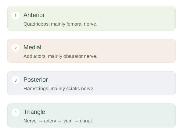
Strong connective tissue divides the thigh into anterior, medial and posterior compartments. Most anterior thigh muscles receive the femoral nerve; the quadriceps straighten the knee. Medial muscles mainly bring the thigh towards the midline and are largely supplied by the obturator nerve. Posterior muscles include the hamstrings, which commonly bend the knee and extend the hip. Most receive the tibial division of the sciatic nerve. These are organising patterns rather than rules without exceptions: individual muscles can have different actions or more than one nerve supply.
Thigh organisation
 Open diagram larger ↗
The femoral triangle
Just below the groin lies a triangular region bounded by the inguinal ligament above, sartorius laterally and adductor longus medially. It contains important structures travelling between abdomen and lower limb. From lateral to medial, the main arrangement is femoral nerve, artery and vein, followed by the femoral canal and lymphatic structures. The nerve lies outside the femoral sheath; the vessels lie within it. This distinction matters because a connective-tissue sleeve does not surround every structure passing through the same visible region.
Routes explain relationships
The femoral artery supplies the limb and gives a large deep branch, the , to the thigh. The femoral vein returns blood towards the pelvis. The nearby femoral canal normally accommodates loose tissue and lymphatics; it is also a potential site of a femoral hernia. Learning the triangle as a map is more useful than memorising its name alone. Imagine a junction where muscles create boundaries and nerves, vessels and lymphatic pathways pass between them. Their proximity explains why clinicians use careful anatomical landmarks when examining a pulse or accessing a vessel.