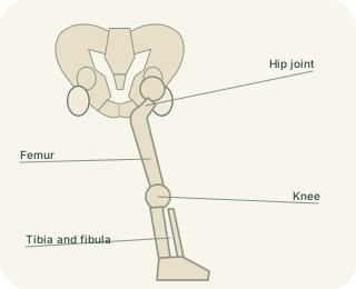
A stable mortise
The ankle joint forms where the lower tibia and fibula surround the upper . This arrangement resembles a socket or mortise holding a moving block. The tibial and fibular malleoli form the bony projections on either side. The talus transfers load down towards the heel and forward through the foot. Its upper surface is wider at the front, so the joint generally fits more tightly in dorsiflexion. Ligaments between tibia and fibula maintain the mortise; these distal connections are called the .
Ankle and hindfoot movements
 Open diagram larger ↗
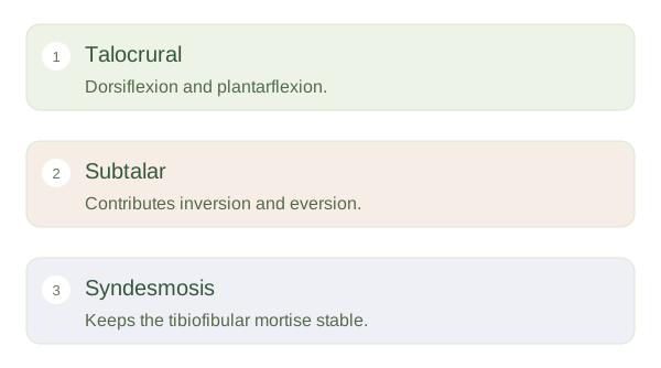
Which joint makes which movement?
The talocrural joint mainly permits dorsiflexion and plantarflexion. Tibialis anterior helps lift the forefoot, while the calf muscles pull through the calcaneal tendon to raise the heel. Inversion and eversion occur mainly through the subtalar and transverse tarsal joints beneath and in front of the . Everyday movements usually combine several joints, which is why rolling a foot cannot be understood as a pure ankle hinge action. Keeping these movements separate in a mental map helps connect muscle actions to the surfaces they move.
Ligaments guide the limits
A strong medial deltoid ligament resists excessive eversion. Lateral ligaments include the anterior talofibular ligament, often stressed when a plantarflexed foot inverts. The exact injury depends on direction, position and force; no single mechanism describes every ankle sprain. Stability also relies on tendon control, sensation and balance responses. When standing on uneven ground, the nervous system adjusts muscle activity continuously. The ankle therefore combines passive guidance from bone and ligaments with active adjustment from muscles. A useful question is always which joint moved and which tissue resisted that movement.