An organised foundation
The foot contains tarsal bones near the ankle, metatarsals in the forefoot and phalanges in the toes. The talus receives load from the leg and passes it towards the calcaneus and the rest of the foot. The calcaneus forms the heel. Longitudinal arches run from heel towards forefoot, while a transverse arch spans across the foot. Their shapes mean the sole is not a flat stack of bones. They help the foot accommodate ground surfaces and distribute forces over the changing areas of contact.
From flexible platform to lever
 Open diagram larger ↗
The arch is not held by bone alone
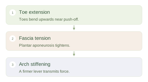
The medial longitudinal arch includes the talus, calcaneus, navicular, cuneiforms and medial metatarsals. Its support comes from joint shape, ligaments, the and muscles. The plantar aponeurosis is a strong sheet beneath the sole, often called plantar fascia. When the toes extend near push-off, this sheet tightens around the heads, helping raise and stiffen the arch. This changes the foot from a more adaptable platform into a firmer lever for propulsion.
Shape and function vary
Tibialis posterior and other extrinsic muscles contribute dynamic support; small intrinsic muscles help control the arch locally. No single tendon carries the entire responsibility. Foot posture varies between people and also changes during movement. A low visible arch does not automatically mean disease, and a high arch does not guarantee effective function. Symptoms and movement matter alongside appearance. Imagine an adjustable bridge supported by several cables: each tissue contributes, and loading changes their tension. This explains why studying only the bones misses much of the foot’s mechanical behaviour during standing, walking and running.