Two sources of muscle control
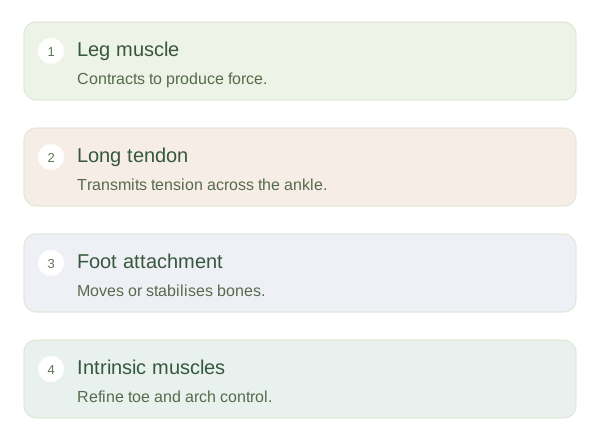
Extrinsic foot muscles begin in the leg and send long tendons across the ankle into the foot. These tendons can move the ankle, support arches and bend or straighten the toes. Intrinsic muscles begin and end within the foot itself. Their shorter lever systems help control toe position and stabilise local joints. Both groups work together: the larger muscles supply much of the force, while smaller muscles adjust how that force reaches the ground. Neither group operates as an isolated replacement for the other.
How force reaches the foot
 Open diagram larger ↗
Following the tendons
The calcaneal tendon joins the calf muscles to the heel and transmits plantarflexion force. Tibialis posterior passes behind the medial malleolus towards the midfoot, contributing inversion and arch support. Fibularis longus passes behind the lateral malleolus and crosses beneath the foot towards the medial forefoot. Long toe flexors travel behind the medial ankle, whereas extensor tendons cross the front. Retinacula are connective-tissue bands that hold tendons close to the skeleton, helping prevent them from bowstringing away from the joint during movement.
Small muscles, meaningful jobs
Plantar interossei and dorsal interossei help control toe alignment; lumbricals coordinate flexion and extension at different toe joints. These actions matter for maintaining contact and balance rather than performing elaborate independent movements like the fingers. Tendons move within sheaths in several locations, which reduces friction as they glide. A tendon can transmit force but cannot shorten actively like muscle. Understanding this distinction makes movement easier to visualise: the muscle contracts at one location, its tendon carries tension along a route and the final attachment changes the position or stability of a bone.