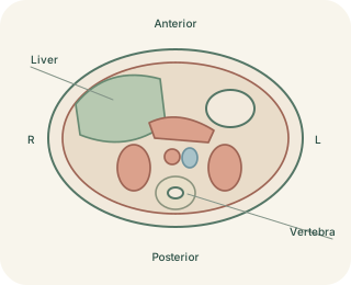
Slices through a three-dimensional body
Cross-sectional imaging displays a thin portion of the body rather than every structure superimposed at once. An axial image cuts across the body, a coronal image divides front from back and a sagittal image divides left from right. These planes are reference directions, not separate anatomical compartments. Imagine slicing a loaf in different directions: the ingredients stay in the same three-dimensional arrangement, but each slice exposes a different pattern. Structures can appear, disappear or change shape as the image moves through them.
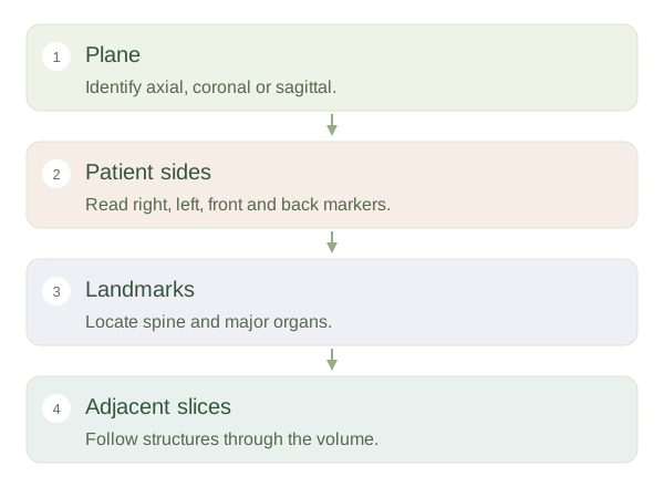
Orient before identifying
 Open diagram larger ↗
The patient sets the directions
Right and left always refer to the patient. Standard axial CT images are usually displayed as though you are looking from the patient’s feet towards the head. The patient’s right then appears on the viewer’s left. The front is normally at the top of the display and the back below. However, labels and orientation markers must be checked rather than assumed. Coronal images commonly resemble facing the patient, while sagittal views require attention to which side and direction are shown. Image acquisition and display settings can vary.
Use relationships before details
Start with reliable landmarks such as the spine, large airways or major vessels, then identify adjacent organs. On abdominal axial images, the liver is mainly on the patient’s right and the spleen on the left. Their presence also depends on slice height. A single cross-section is therefore an incomplete sample of an organ. Following neighbouring slices helps distinguish a continuous tube from an isolated round structure. Beginner image reading should build orientation and spatial understanding first; recognising a landmark is different from making a diagnosis or excluding disease from one selected picture.