From source to detector
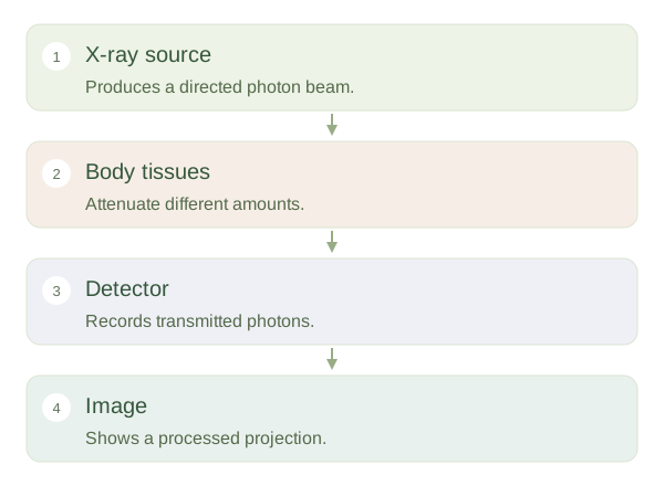
An x-ray machine sends a controlled beam of high-energy photons through the body towards a detector. Some photons pass through; others are absorbed or scattered. This reduction of the original beam is called . Different tissues attenuate the beam differently according to factors including their thickness, composition and density. The detector records the photons that arrive, and the system converts their distribution into an image. The photograph-like appearance can be misleading: it represents transmission through the body rather than light reflected from its surface.
Making a radiograph
 Open diagram larger ↗
Why bone looks white
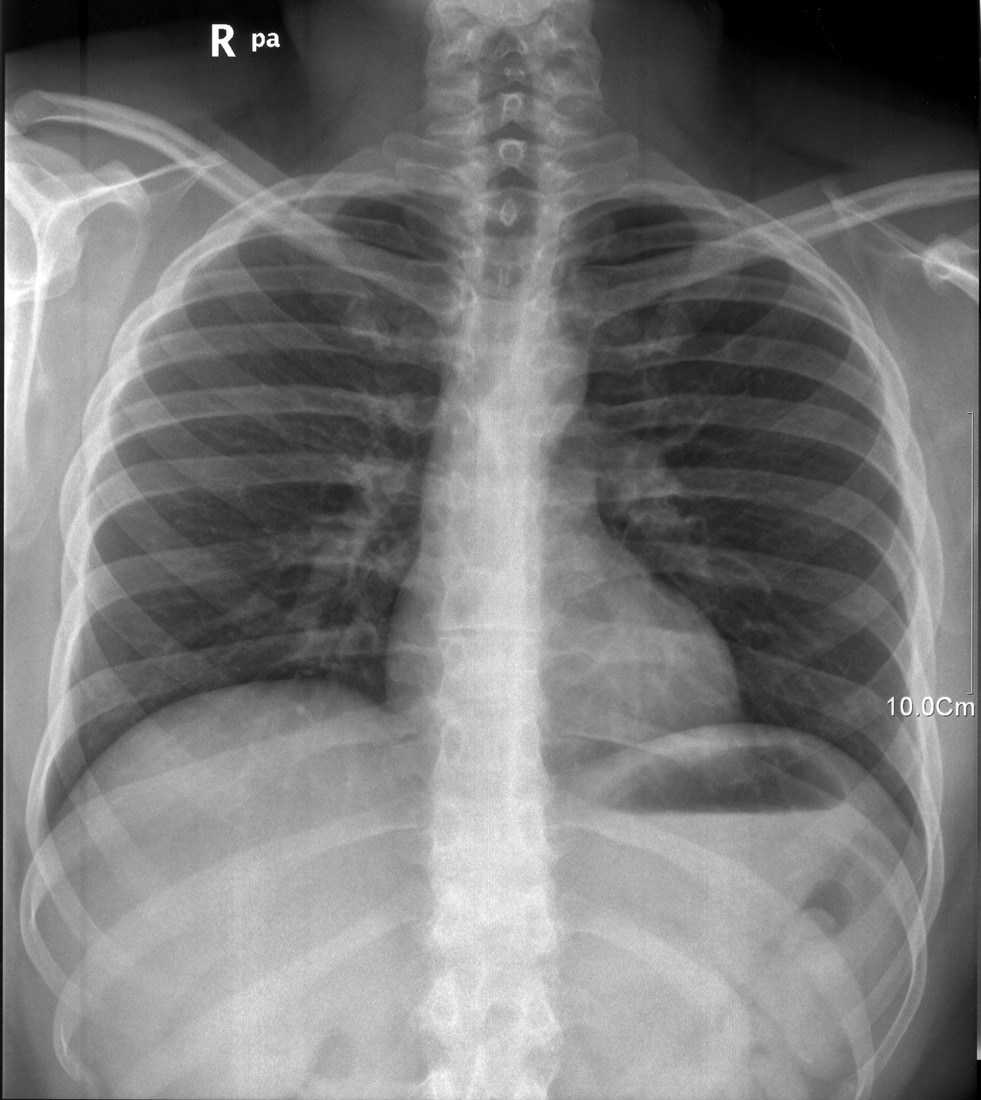
On a conventional radiograph, strongly attenuating material generally appears lighter because fewer photons reach the detector behind it. Bone and many metals are therefore bright. Air attenuates much less and appears dark; soft tissues occupy intermediate shades. A thicker region can appear denser even when its material is unchanged, because the beam passes through more tissue. The displayed brightness also depends on processing settings. A white patch does not identify one substance by itself: bone, overlapping soft tissue, fluid and other materials can all affect the appearance.
One picture contains overlap
A plain radiograph compresses three-dimensional anatomy into a two-dimensional . Ribs, lungs, heart and skin can contribute to the same part of a chest image. A second view from a different direction can help separate structures. This is one reason an apparent finding must be interpreted with positioning, technique and clinical information. X-rays are ionising radiation, so examinations use justified exposures and methods that limit dose while preserving useful detail. No radiation remains in the body after an ordinary x-ray exposure. The resulting picture can be stored and reviewed without exposing the patient again.