A probe that sends and listens
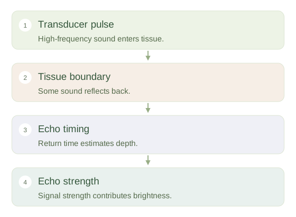
An ultrasound contains elements that convert electrical energy into high-frequency sound and returning sound into electrical signals. The probe sends short pulses into the body, then listens for echoes. Sound reflects partly where tissues with different acoustic properties meet. The machine estimates depth from the time taken for an echo to return, using an assumed speed of sound in soft tissue. Echo strength helps determine brightness. Repeating this across many beam directions produces a sectional image that can update rapidly enough to show movement.
Pulse, echo, image
 Open diagram larger ↗
What creates a good acoustic window?
Gel removes the air gap between probe and skin because air strongly disrupts sound transmission. Fluid often transmits sound well, whereas bone and gas can block or scatter it and hide deeper structures. Higher-frequency sound generally gives better detail near the surface but loses strength more quickly with depth. Lower frequencies penetrate further with less fine detail. Probe choice and position therefore depend on the organ and the question. Ultrasound does not reveal everything equally well simply because the probe is touching the body.
Real time, with limitations
Ultrasound can show a beating heart, moving tendons or changes during breathing. The person holding the probe chooses the plane actively, so anatomy may be displayed obliquely rather than in a standard textbook section. Some apparent features are artefacts produced by how sound travels, such as shadows behind strongly attenuating material. Ultrasound uses non-ionising sound rather than x-rays, but sensible examination settings and trained use still matter. The central idea is echo location: the machine maps returning signals, and the image must be interpreted with the beam path and surrounding anatomy in mind.