Moving cells change the echo
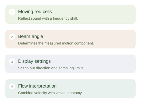
When ultrasound reflects from moving red blood cells, the returning frequency differs slightly from the transmitted frequency. This provides information about motion relative to the probe. The size and direction of the shift depend on blood speed and the angle between flow and the beam. Flow directly towards or away from the probe produces a larger useful shift than flow across the beam. Near a right angle, conventional Doppler measurements become unreliable because little motion lies along the beam direction.
Reading a Doppler signal
 Open diagram larger ↗
Several ways to display flow
Colour Doppler overlays a map of detected motion on a grey-scale anatomical image. A common convention shows flow towards the probe in one colour and away in another, but the map can be inverted. Red does not inherently mean oxygenated blood or artery, and blue does not inherently mean vein. plots measured velocities over time, revealing pulsation and flow patterns. Power Doppler emphasises signal strength and can help detect slow flow, but standard power displays do not provide the same directional information.
A measurement with conditions
A narrowed vessel may produce locally increased velocity, but interpretation requires correct angle settings, sampling position and the wider pattern. Technical limits can make a normal flow appear absent. occurs when measured shifts exceed the selected sampling range, producing a wrapped display rather than a literal reversal of blood. Motion of the probe or surrounding tissue can also introduce signals. The useful habit is to read the colour scale and the measurement settings before interpreting the picture. Doppler adds functional information to anatomy; it does not turn every colour patch into a diagnosis on its own.