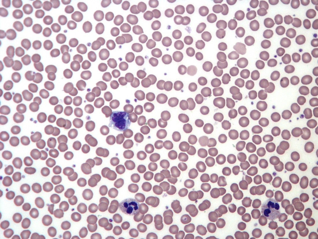
Spreading blood into a thin layer
A is made by spreading a small blood sample across a slide and staining it. In a suitable region, cells lie mostly separately rather than piled on top of one another. This allows their size, shape and internal features to be compared. The image shows a sample of circulating components, not all the blood or every immune cell in the body. The stain introduces colours to reveal structures; the colours are not identical to what an unstained cell looks like in a living vessel.
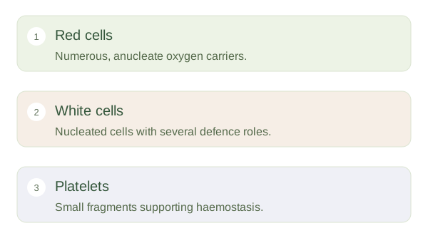
Three components of a blood film
 Open diagram larger ↗
The main components
Red blood cells dominate a normal field. Mature human red cells have no nucleus and commonly show a paler centre reflecting their biconcave shape. White blood cells are much fewer and have nuclei. Neutrophils often have segmented nuclei; lymphocytes commonly show a large rounded nucleus relative to their cytoplasm. Monocytes are usually larger with more folded nuclei. Eosinophils and basophils have distinctive granules on suitable stains. Platelets are small cell fragments involved in haemostasis. They are not miniature nucleated white cells, even though they can appear as tiny coloured dots.
Pattern before diagnosis
A smear can reveal variation in cell size, shape, maturity or distribution. Red-cell appearance can help investigate anaemia, while white-cell patterns can guide further questions about infection or blood disorders. However, one unusual cell or selected photograph rarely establishes the whole diagnosis. Preparation can create clumps, damaged cells or uneven distribution. A full blood count supplies numerical information that complements , and additional tests may be needed. First learn to distinguish red cells, white cells and platelets, then relate their structures to oxygen transport, defence and clotting rather than treating every visible difference as disease.