A shared ability to generate force
All three muscle types use specialised proteins to produce force, but they arrange those proteins and their cells differently. Skeletal muscle moves the skeleton and contributes to posture and heat production. Cardiac muscle forms the pumping wall of the heart. Smooth muscle controls many hollow organs and blood vessels. Microscopic classification connects shape with function, yet it does not mean every action is either completely voluntary or completely automatic. Skeletal muscle, for example, also participates in reflexes and involuntary shivering.
Muscle tissue comparison
 Open diagram larger ↗
Reading the structural differences
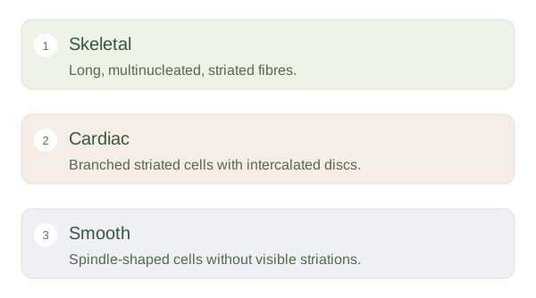
Skeletal fibres are long cylindrical cells with many nuclei, usually located near the cell surface. Their regular protein arrangement produces visible striations in suitable longitudinal sections. Cardiac cells also have striations but are shorter and branching, typically with one central nucleus. Intercalated discs join neighbouring cardiac cells mechanically and electrically. Smooth muscle cells are , usually have one central nucleus and lack obvious cross-striations on routine light microscopy. Their contractile proteins are present but are not arranged in the repeating sarcomere pattern that creates striations.
Orientation can hide the clues
In cross-section, skeletal fibres look like rounded or polygonal profiles rather than long bands. Not every section through a smooth muscle cell includes its nucleus, because the section may miss the wider central region. A poorly oriented cardiac section may not show clear intercalated discs. Use several features and the organ context together rather than relying on one missing line. The differences support different jobs: skeletal muscle can generate directed movement, cardiac cells coordinate a repetitive pump and smooth muscle sustains or changes tension in organ walls. The tissue’s organisation is part of its physiological strategy.