More than neurons alone
Nervous tissue contains neurons, which process and transmit signals, and glial cells, which support and regulate their environment. A neuron’s cell body contains its nucleus and much of its protein-making machinery. Dendrites commonly receive inputs, while an axon carries signals towards other cells. have varied roles, including maintaining local conditions, participating in immune defence and forming . A routine slide shows only part of this network. Many fine processes and synapses are too small or too weakly contrasted to follow completely with ordinary light microscopy.
Nervous tissue organisation
 Open diagram larger ↗
Grey matter, white matter and nerves
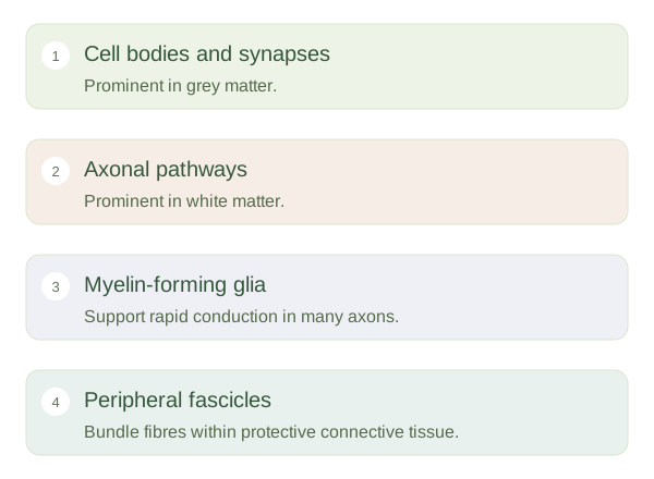
Grey matter contains many neuronal cell bodies, dendrites, synapses and supporting cells. White matter contains many axons, often wrapped in . The distinction is relative: neither region contains only one kind of structure. Myelin forms from oligodendrocytes in the central nervous system and Schwann cells around peripheral axons. In a peripheral nerve, fibres are bundled into fascicles surrounded by connective-tissue layers. A nerve therefore differs from a neuron: it is a larger organised pathway containing many axons and associated supporting structures.
The preparation influences the picture
contains substantial lipid, and routine processing can remove some of it, leaving pale spaces around axons. Special stains can reveal components that ordinary H&E shows poorly. A cross-section of a nerve looks very different from a longitudinal section, just as a cable looks different when cut across its wires. Large cell bodies can suggest neuronal regions, but exact cell identification may require additional methods. The practical goal is to distinguish cell bodies, fibre-rich areas and supporting organisation, then relate them to local processing or long-distance communication without assuming the slide displays every functional connection.