From tissue to working organ
The four basic tissue families are epithelial, connective, muscle and nervous tissue. An organ combines several of them in a particular arrangement to perform a coordinated task. The stomach, for example, is not simply a muscular bag. Its lining must manage acid and secretion, its supporting tissues carry vessels and nerves, and its muscle layers mix and move contents. Looking at a whole wall provides a different level of explanation from identifying an isolated cell. Structure becomes useful when it is connected to the organ’s overall job.
A coordinated organ wall
 Open diagram larger ↗
Follow a hollow organ wall
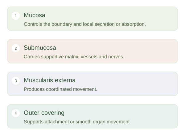
In much of the digestive tract, the includes an epithelial boundary and supporting tissue. Beneath it, the contains connective tissue with vessels and nerves. The provides larger muscle layers that move contents, with neural networks coordinating activity. An outer covering attaches the organ or allows movement against nearby surfaces, depending on its location. The exact layers and specialisations vary along the tract. The intestine needs substantial absorptive surface, while the oesophagus needs protection from abrasion. The shared plan is adapted rather than copied identically.
Supply and communication connect the parts
Blood vessels deliver oxygen and nutrients, nerves regulate activity and connective tissue transmits forces between layers. The epithelium controls what crosses the boundary, but it depends on those supporting systems. Damage to one component can disturb the whole organ: reduced blood supply can impair the lining and muscle together, while abnormal movement can affect digestion despite intact secretory cells. When reading a slide, first identify the lumen or surface, then work through the layers and their relationships. The question is not only what tissue is present, but how its position enables cooperation.