A clot in the returning circulation
Deep vein thrombosis, or DVT, is a clot forming in a deep vein, commonly in the lower limb or pelvis. Venous clotting is favoured by slowed flow, vessel-wall injury and an increased tendency of blood to clot. These factors can occur in many combinations, including immobility, surgery, illness or inherited influences. A may obstruct local return and provoke swelling or pain, although signs can be subtle or absent. A clot in a deep vein is different from an atherosclerotic plaque developing in an arterial wall.
From deep vein to pulmonary artery
 Open diagram larger ↗
The route to the lungs
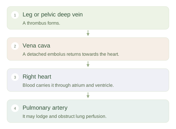
Part of a can detach and travel as an . Venous blood from the lower body flows through the inferior vena cava into the right atrium, right ventricle and pulmonary arteries. A clot following this route may lodge in a pulmonary artery, producing a . The vessel is called an artery because it carries blood away from the heart, even though this blood has not yet passed through the lungs for oxygenation. Normal circulation therefore explains why a leg clot usually threatens the lungs rather than travelling directly to the brain.
Two consequences of obstruction
An affected lung region may still receive air while receiving too little blood, reducing effective matching of ventilation and perfusion. A large or widespread obstruction can also increase the load on the right ventricle. Severity depends on the amount and location of obstruction and the person’s existing cardiopulmonary reserve. Not every DVT produces an embolism, and symptoms alone do not confirm either diagnosis. Venous thromboembolism is the umbrella term covering both conditions. The important anatomical lesson is to trace the route: the consequences of a travelling clot depend on where it enters circulation and which vessels lie downstream.