Slicing a three-dimensional body
A body is three-dimensional, but most textbook sections and many medical images are flat. Anatomical planes provide a shared way to describe the direction of a slice. Think of slicing a loaf: a change in cutting direction produces a very different outline, even though the loaf is unchanged. A plane is an imaginary flat surface; a section is the view produced along it. Seeing an unusual shape on one section does not automatically mean an organ itself has an unusual shape.
Anatomical Planes and Sections
 Open diagram larger ↗
The three main directions
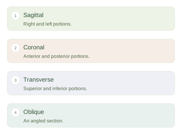
A divides right from left. A midsagittal section passes through the midline and creates right and left halves; other sagittal sections are off-centre. A frontal, or coronal, plane divides anterior from posterior portions. A cuts across the body, dividing superior from inferior portions. A transverse section through the trunk resembles looking at a slice across a cylinder, although real anatomy is much more irregular. Oblique sections cross the body at an angle rather than following one of these principal directions.
Reading sections carefully
One slice shows only structures that meet that particular level. A transverse view through the chest cannot show every part of both lungs, the whole heart and the entire diaphragm simultaneously. Following neighbouring slices helps reveal how structures continue through space. The direction from which an image is viewed also matters: standard radiological displays often show transverse images as if looking from the feet towards the head. Check orientation labels rather than guessing. First identify the plane, then the level, and finally the structures within that section.