Injury starts a repair response
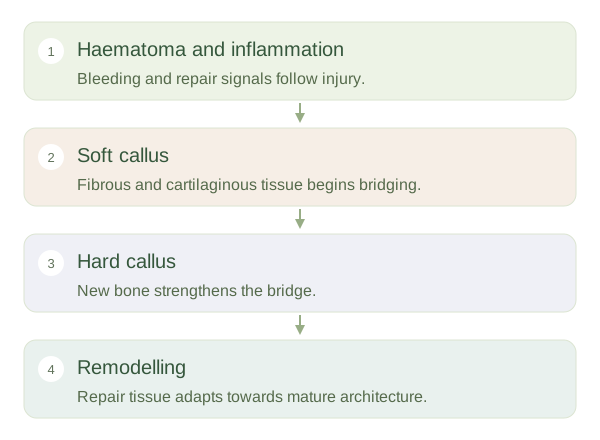
A disrupts bone tissue and often nearby vessels. Bleeding creates a around the break, and inflammatory signals recruit cells that clear damaged material and begin repair. This early stage is biologically active, even though no new solid bridge is yet visible. The periosteum, marrow and surrounding tissues contribute cells and blood supply. Stability and adequate circulation are important because repair tissue must form across the injured region. Infection, major soft-tissue damage or poor blood supply can interfere with the process.
How Fractures Heal
 Open diagram larger ↗
A bridge forms
In the common pattern of secondary healing, a soft develops from fibrous tissue and cartilage around the break. It provides an initial bridge but is not as strong as mature bone. New bone gradually replaces much of this tissue, producing a hard bony callus. The process resembles aspects of developmental bone formation but occurs within an injury environment. Not every fracture heals through an identical sequence: very stable, closely opposed fractures may heal more directly with less obvious callus. The mechanical conditions influence which repair route predominates.
Remodelling takes longer
After the is bridged, osteoclasts and osteoblasts continue reshaping the repair. Trabeculae and the outer contour adapt to mechanical demands, and excess may be removed. Functional recovery therefore does not mean the tissue has instantly regained its original architecture. Healing speed varies with the bone, injury, age and health context. The key concept is a coordinated progression from injury response to temporary support, bony bridging and later . This is a biological explanation, not a guide to setting a fracture. It shows why a broken bone needs both living repair capacity and an appropriate mechanical environment, rather than simply waiting for mineral to glue the pieces together.