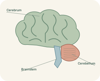
A folded outer processing layer
The cerebrum forms the largest part of the brain. Its outer cortex is a folded sheet containing many neuronal cell bodies. Folding increases the amount of cortical surface within the skull. Each hemisphere is divided into major lobes using visible grooves as landmarks. These boundaries help describe anatomy, but they are not sealed compartments. Nerve fibres link nearby and distant regions, including connections between the two hemispheres through the .
Cerebral lobes specialise, but everyday tasks depend on connected networks.
 Open diagram larger ↗
Four useful landmarks
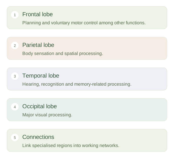
The frontal contributes to planning, decision-making, behaviour and voluntary movement. Its primary motor cortex lies near the central . The parietal lobe helps process body sensation and spatial relationships; its primary somatosensory cortex lies just behind that groove. The occipital lobe contains major visual-processing areas. The temporal lobe contributes to hearing, recognition, memory and aspects of language. Deeper within the lateral sulcus, the insula participates in bodily awareness and other functions. These are starting points rather than complete descriptions of each region.
A network view is more accurate
Reading a sentence uses visual regions to analyse symbols, language networks to interpret meaning, memory systems to supply context and attention networks to maintain focus. No single does the whole job. Some functions are more strongly represented in one hemisphere, such as language in the left hemisphere for many people, but this varies. Most people do not have a purely logical hemisphere and a purely creative one. Motor and sensory pathways also often link one cerebral hemisphere with the opposite side of the body. A brain injury’s effects depend on the specific region, connections and extent involved, rather than simply the lobe’s name. Combining regional maps with an understanding of networks makes cerebral anatomy much more useful than memorising a list of isolated functions.