A tube with several jobs
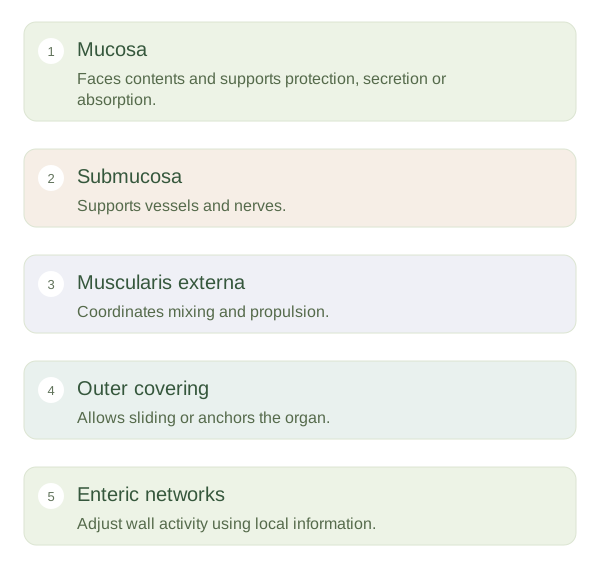
The digestive tract is a continuous tube from mouth to anus. Material inside its has not yet crossed into the body’s internal tissues. Much of the tract has a shared wall plan. The faces the contents and includes an epithelial lining adapted for protection, secretion or absorption. Beneath it, supporting tissue contains vessels and immune cells. The submucosa provides another supportive layer carrying larger vessels, nerves and other structures.
The gut wall combines a barrier, transport surface and coordinated muscle.
 Open diagram larger ↗
Muscle moves and mixes
The muscularis externa usually contains circular and longitudinal smooth muscle layers. Coordinated contraction can propel material along the tube, a pattern called . Other patterns mix contents, improving contact with digestive secretions and absorptive surfaces. The arrangement varies by region: the stomach has additional muscle organisation for churning, while swallowing and defaecation involve some skeletal muscles. The outer covering may be a slippery serosa or an anchoring adventitia, depending on the organ’s relationship to surrounding tissues.
A local nervous system
Enteric nerve networks within the wall help control movement, secretion and local blood flow. They can organise many responses without a separate conscious command for every contraction. Autonomic signals, hormones and conditions inside the gut modify this activity. Stretch from a meal and the chemical composition of contents provide information about what the gut needs to do next. The system is therefore locally capable but not disconnected from the rest of the body. A useful analogy is a transport line with its own sensors and controllers, while central management still influences the overall schedule. Understanding this shared wall plan makes differences between the oesophagus, stomach and intestine easier to learn: each adapts the same basic architecture to a different stage of digestion.