Meeting in the uterine tube
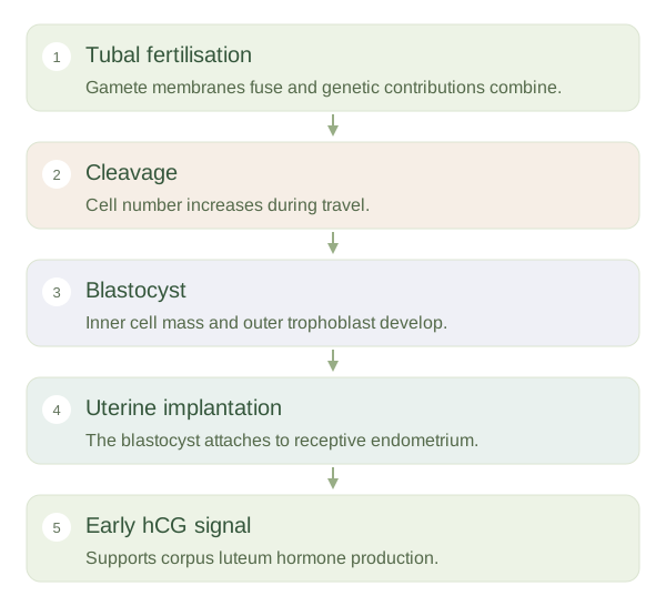
Fertilisation is the process by which a sperm and an oocyte combine their genetic contributions. It usually occurs in the ampulla of a uterine tube. Sperm must first undergo functional changes called capacitation in the reproductive tract. Interactions with the oocyte's surrounding layers help prepare a sperm to fuse with its membrane. Fusion normally triggers completion of the oocyte's second meiotic division and changes that reduce entry by additional sperm.
Fertilisation is not implantation
 Open diagram larger ↗
Dividing without immediately growing
The resulting begins : repeated cell divisions that increase cell number while the overall structure initially remains within its surrounding coat. It travels towards the uterus with assistance from tubal cilia and muscular activity. A compact cell cluster develops into a containing an inner cell mass and an outer trophoblast. The inner group contributes to the embryo, while trophoblast tissue contributes to supporting structures, including the fetal part of the placenta.
Attaching to the lining
begins when the attaches to and enters a receptive endometrium, usually starting about a week after fertilisation. The outer cells interact closely with maternal tissue and begin establishing an exchange interface. They also produce hCG, which helps maintain the corpus luteum and its hormone support of the endometrium. Fertilisation and implantation are therefore separated by development, transport and time, rather than being two names for the same event.
Location matters
Successful development depends on the embryo and the uterine lining being suitably coordinated. outside the normal uterine cavity is called an ectopic pregnancy, most commonly involving a uterine tube. This shows why a tube is suited to early transport but not to supporting an expanding pregnancy. Many early developmental events do not lead to an ongoing pregnancy. This article describes the usual biological sequence without assuming that every fertilised oocyte will implant or develop normally.