A protective frame that can move
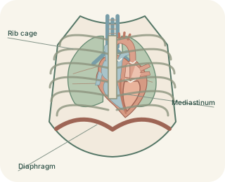
The thoracic cage is formed by thoracic vertebrae, ribs, costal cartilages and the . There are usually twelve pairs of ribs. The first seven pairs attach to the sternum through their own costal cartilages. Ribs eight to ten connect indirectly through cartilage above, while the final two pairs have no anterior attachment to the sternum. The sternum consists of the manubrium, body and xiphoid process. This arrangement protects the heart and lungs while allowing changes in chest dimensions during breathing.
The chest wall as a functional frame
 Open diagram larger ↗
What lies between the ribs
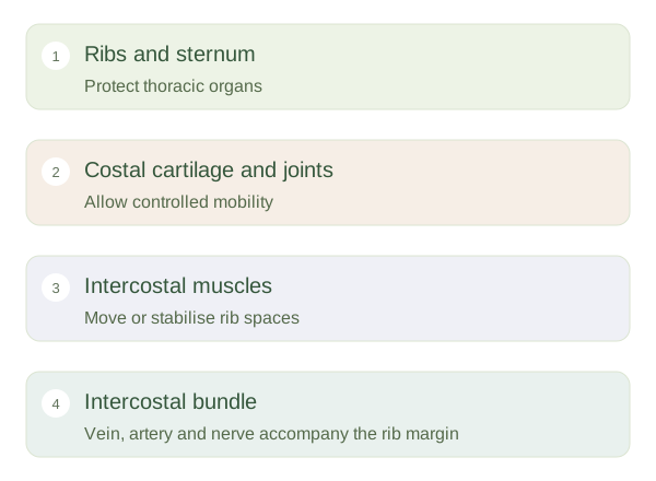
Intercostal spaces contain layers of muscle, nerves and blood vessels. External, internal and innermost intercostal muscles have different fibre directions and contribute to movement or stabilisation according to the task. Quiet inspiration mainly depends on the diaphragm, with rib movement assisting expansion. Forced breathing recruits additional muscles. The main intercostal runs near the inferior border of each rib, between internal and innermost muscle layers. Its usual superior-to-inferior order is vein, artery and nerve, although smaller collateral branches and anatomical variation are important too.
Movement changes volume
Upper rib movement can increase the front-to-back dimension of the chest, while more lateral movement increases its width. These are often described as pump-handle and bucket-handle patterns, useful analogies rather than perfectly separate mechanisms. provides flexibility, and joints at the vertebral end permit controlled motion. A painful chest-wall injury may therefore affect breathing without directly damaging lung tissue. Regional anatomy links the rigid-looking bones with the mobile joints and soft tissues between them. Protection and ventilation are compatible because the cage is strong without being a single immovable shell.