Glandular tissue within a soft framework
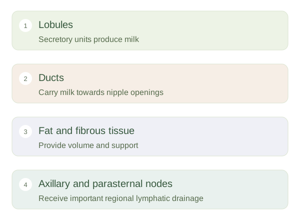
Breast tissue lies in the superficial tissues of the anterior chest, largely over pectoralis major. It includes glandular structures, fat, fibrous support, vessels and nerves. Milk-producing units called lobules connect with ducts leading towards openings at the nipple. The surrounding pigmented skin is the . Fibrous bands support the tissue and link it with surrounding structures, while a loose plane helps the breast move over the deeper muscle. The amount of glandular tissue and fat varies between individuals and across life stages.
Breast structure and lymphatic connections
 Open diagram larger ↗
Function changes across life
Hormones influence breast development and changes during menstrual cycles, pregnancy and lactation. During lactation, secretory cells produce milk, and surrounding myoepithelial cells contract in response to oxytocin to help move it into ducts. Milk production and milk ejection are related but different processes. Breast size alone does not reliably indicate milk-producing capacity because size also reflects fat and other tissue. All sexes can have breast tissue, although its usual development and hormonal responsiveness differ. The regional anatomy should therefore be understood through tissue composition and function rather than appearance alone.
Lymphatic routes explain connections
Lymph vessels collect excess tissue fluid and carry it through regional nodes. Much breast lymph drains towards axillary nodes in the armpit, while medial regions also communicate with parasternal nodes near the sternum. Drainage patterns vary and can cross expected boundaries. These routes are clinically relevant because some cancer cells may travel along lymphatic pathways, but enlarged nodes do not by themselves prove cancer. A sentinel node is an early draining node identified for a particular region, rather than a single fixed node in every person. The useful concept is a connected tissue-fluid network, not isolated breast tissue with only one exit.