An oblique organ in the central chest
The heart lies within the middle mediastinum, behind the sternum and between the lungs. It is not entirely on the left: a substantial portion lies near or to the right of the midline. Its pointed apex usually faces downwards, forwards and towards the left, while its broader base faces mainly backwards. The right ventricle forms much of the anterior surface, and the left atrium contributes much of the posterior base. These relationships help translate a familiar four-chamber diagram into the body’s three-dimensional arrangement.
Heart surface to surrounding sac
 Open diagram larger ↗
A sac that permits movement
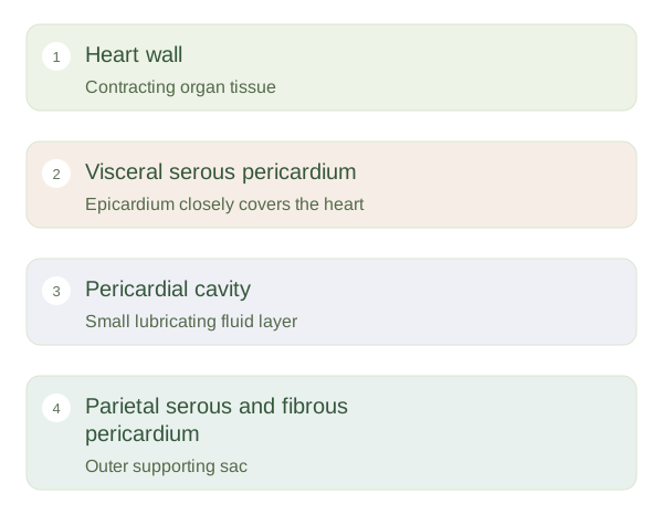
The pericardium surrounds the heart and the roots of its major vessels. Its fibrous outer component helps support position, while serous layers provide smooth surfaces. The visceral serous layer closely covers the heart and is also called the . A small amount of fluid normally lies between visceral and parietal serous layers, reducing friction as the heart moves. The heart is not floating inside a large fluid-filled chamber. Excess fluid can become important if it raises surrounding pressure enough to impair filling, depending partly on how quickly it accumulates.
Connections at the top and back
The aorta carries blood from the left ventricle into the systemic circulation. The leaves the right ventricle and divides towards the lungs. Superior and inferior venae cavae return systemic blood to the right atrium, while pulmonary veins return oxygenated blood to the left atrium. These great vessels anchor the organ while allowing rhythmic motion. Coronary vessels serve the heart muscle itself rather than replacing these main inflow and outflow routes. Regional anatomy combines chamber orientation, the surrounding sac and vessel connections into one coherent map of the beating heart.