A suspension system, not one shelf
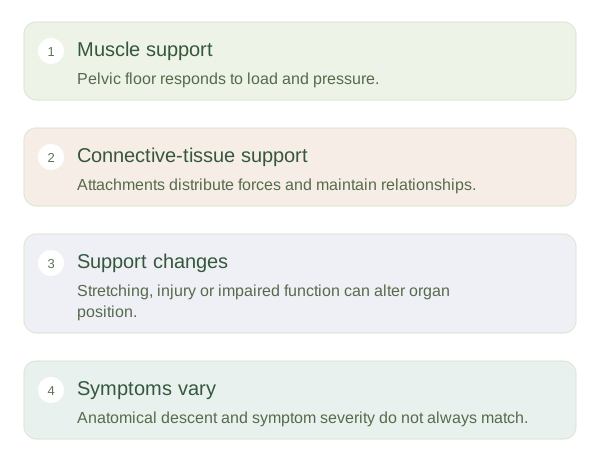
Pelvic organs are supported by the pelvic floor muscles, connective-tissue attachments and surrounding structures. The supports distribute forces during standing, lifting, breathing and changes in organ filling. They do not hold each organ independently in a rigid box. When part of this combined system stretches or loses function, organ position may change. In , vaginal walls, the uterus or the vaginal apex can descend. The bladder or rectum may bulge with the adjacent vaginal wall. The process usually reflects interacting support changes rather than a single structure suddenly detaching.
Pelvic Support Changes and Prolapse — concept map
 Open diagram larger ↗
Different sites, different relationships
An anterior vaginal-wall prolapse may involve the bladder and is often called a . A posterior vaginal-wall bulge may involve the rectum and is often called a . Apical prolapse affects the uterus or the top of the vagina. These descriptions identify the affected compartment but do not by themselves tell us how severe symptoms will be. Rectal prolapse, where rectal tissue protrudes through the anus, is a different condition even though both involve support and descent. Understanding the route of the bulge is therefore as important as knowing which organ is nearby.
Why anatomy and symptoms can differ
Pregnancy and childbirth, ageing, connective-tissue characteristics, surgery and repeated pressure loads can influence support. They are risk factors, not guarantees that prolapse will occur. A person may have an anatomical change with few symptoms, while another has pressure, a bulge sensation or altered bladder or bowel function. Symptoms require assessment in context because similar complaints can have other causes. The anatomical model explains why management considers the entire support system and the person’s experience rather than an image alone. This article does not prescribe exercises or treatment: coordinated relaxation, tissue support and nerve function matter alongside muscle strength.