A triangular front-of-elbow space
The is a triangular region on the anterior side of the elbow. Brachioradialis forms its lateral boundary and pronator teres its medial boundary. An imaginary line between the humeral epicondyles forms its upper base. Brachialis and supinator contribute to the floor, while skin, fascia and the lie above the deeper contents. This organisation matters because visible veins near the skin are not in the same layer as every important structure underneath. A surface landmark helps locate a region but does not reveal its full depth.
Cubital Fossa and Important Structures — concept map
 Open diagram larger ↗
Contents have an order
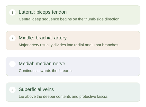
A useful lateral-to-medial sequence for the central deep contents is biceps tendon, brachial artery and median nerve. The radial nerve lies farther laterally, partly under brachioradialis, where it divides into branches. The brachial artery usually divides into radial and ulnar arteries near the distal part of the fossa. The biceps tendon passes towards its radial attachment and helps produce elbow flexion and supination. The median nerve continues into the forearm, carrying fibres to many flexor muscles and sensory territories farther down the limb.
Superficial access, deeper protection
The commonly connects superficial veins across the front of the elbow and is often used for taking blood. Its precise pattern varies. The is a sheet from the biceps tendon that provides some separation between superficial veins and deeper artery and nerve. This is an anatomical explanation, not an instruction for performing a procedure. It illustrates why depth and individual variation matter whenever structures are close together. The acts as a transition zone: a major arm artery branches, nerves continue towards the forearm, and a muscle tendon crosses to its insertion.