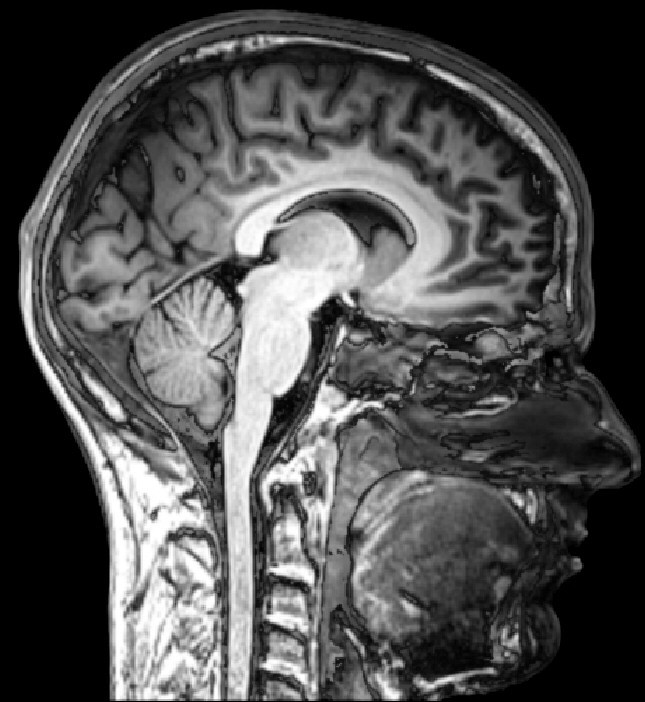
Signals from hydrogen
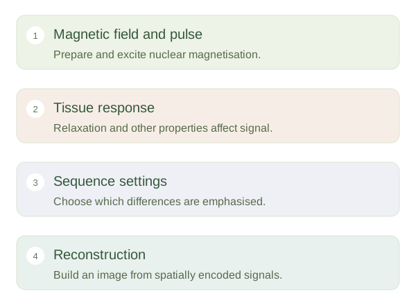
MRI uses a strong magnetic field and radiofrequency pulses to detect signals associated mainly with hydrogen nuclei in body water and fat. After a pulse changes their magnetic behaviour, the nuclei return towards equilibrium and generate measurable signals. Coils detect these signals, and magnetic-field gradients help locate where they arose. A computer reconstructs an image. MRI does not use ionising x-rays. Its image brightness therefore cannot be interpreted as though it were the same attenuation scale used by CT.
MRI contrast depends on the measurement
 Open diagram larger ↗
Sequence changes the contrast
Tissues differ in properties including T1 and T2 , proton density and water movement. A chooses how these differences influence the image. On many images, simple fluid appears bright; on many T1-weighted images, fluid appears dark and fat is relatively bright. Fat suppression and other techniques can deliberately change these patterns. It is therefore unsafe to memorise that water or fat always has one shade on MRI. First identify the sequence, then ask how a tissue should behave under those settings.
Strengths and practical limits
MRI can distinguish many soft tissues and show structures in several planes. Different sequences may reveal complementary information about the same region. Movement can blur images because data are collected over time. Strong magnets also require careful screening of implants, equipment and loose metal objects; an implant is not automatically unsafe, but its specific conditions matter. Some examinations use gadolinium-based contrast to change local signal. A useful mental model is listening to the same orchestra with different microphones: the anatomy remains, but the selected measurement highlights different features. Interpreting several sequences together is more reliable than judging one bright area alone.