The signal begins inside
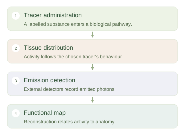
In nuclear medicine, a small quantity of a radioactive substance is linked to a carrier or given in a form that follows a biological pathway. This may be injected, swallowed or inhaled, depending on the test. A detector outside the body records emitted radiation. This differs from an x-ray examination, where an external beam passes through the body. The tracer’s distribution can reveal processes such as tissue metabolism, blood supply or organ handling of a substance, rather than only the outline of an organ.
Following a radiotracer
 Open diagram larger ↗
What PET adds
Positron emission tomography uses tracers that emit positrons. When a positron meets an electron, the interaction produces photons that can be detected in paired directions. Reconstruction estimates where these events arose. A common tracer, fluorodeoxyglucose, behaves similarly to glucose in important ways and can highlight tissues taking it up. Increased is not specific to cancer: normal brain activity, inflammation and other processes may also produce strong signals. Other tracers target different biological properties, so not every scan is a glucose scan.
Function and anatomy together
is often combined with CT to relate tracer activity to detailed anatomy; PET/MRI is another possibility. The anatomical and functional components answer complementary questions. Tracer activity declines through radioactive decay and biological clearance, with timing depending on the substance. The examination therefore has preparation and timing requirements. A bright region needs interpretation with normal tracer distribution, technique and clinical information. Think of the scan as mapping where a labelled participant travels in a biological process, not as a universal detector that marks all disease. The choice of tracer determines which question can be answered.