Not all imaging uses radiation
Plain x-rays, CT and many nuclear-medicine examinations use . This energy can remove electrons from atoms and, in sufficient circumstances, damage biological molecules including DNA. Ultrasound and MRI do not use ionising radiation, although they have their own practical safety considerations. Radiation dose varies greatly by examination, body region, equipment and patient factors. It is therefore misleading to treat every medical image as carrying the same exposure or to compare tests without specifying what is actually performed.
Choosing an imaging examination
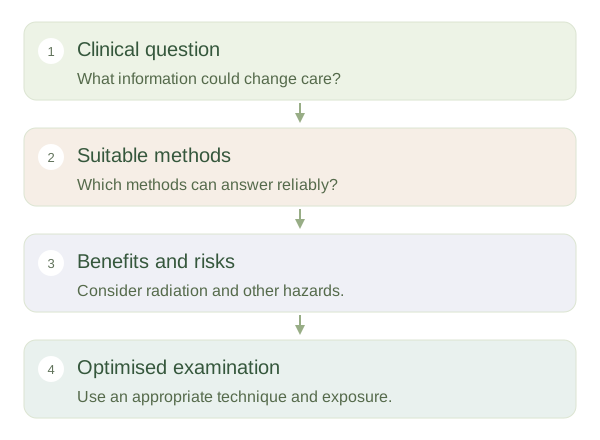 Open diagram larger ↗
Justification and optimisation
A justified examination is expected to provide information useful enough to outweigh its potential harms. aims to obtain an adequate answer using an appropriate exposure, rather than pursuing the prettiest image at any dose. Children, pregnancy and repeated examinations can require particular consideration, but a needed investigation may still have substantial benefit. An image that quickly identifies dangerous internal bleeding, for example, can change immediate care. The decision concerns the whole situation, including alternative tests and the consequences of delaying or missing important information.
Risk is not the same as certainty
A small statistical risk does not mean an individual examination will cause harm, and an absence of immediate symptoms does not mean all risk is zero. Estimates at low doses contain uncertainty. MRI screening focuses on issues such as magnetic interactions with devices and heating; ultrasound settings consider energy delivery and exposure time. Contrast and sedation can add separate risks independent of the image-producing method. Good decisions compare the actual question, useful accuracy, availability and patient circumstances. The educational goal is to understand these categories, not to decide that one modality is always safe or another should always be avoided.