Start with the level
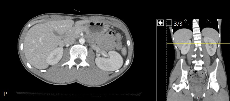
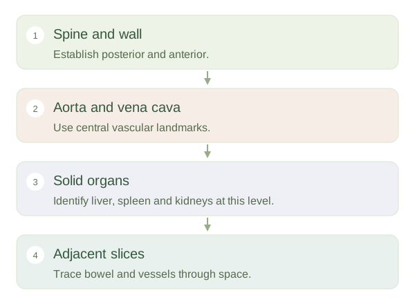
An axial abdominal image shows a horizontal sample through the body. A section near the upper abdomen may include liver, spleen and stomach; lower sections show different parts of the pancreas, kidneys and bowel. An organ is not necessarily missing because it is absent from one selected image. On the conventional display, the patient’s right appears on the viewer’s left. Find the vertebral body at the back and the abdominal wall at the front before searching for smaller structures. Always check orientation labels on the actual study.
Orienting an abdominal slice
 Open diagram larger ↗
Build a map around stable landmarks
The abdominal usually lies just in front of the spine and slightly to the patient’s left. The lies to its right. Kidneys sit posteriorly, outside the main peritoneal cavity, with surrounding fat helping outline them. The liver occupies much of the right upper abdomen, while the spleen lies to the left near the stomach. The pancreas runs across the posterior upper abdomen, behind much of the stomach. These are three-dimensional relationships, so a single slice may intersect only parts of several structures rather than their complete textbook shapes.
Brightness needs context
On CT, gas in bowel is dark, fat is darker than many soft tissues and dense bone is bright. Vessels may become especially conspicuous after contrast injection. Their appearance depends on the acquisition phase and display window. Bowel loops vary in position and contents; identifying every round structure from shape alone is unreliable. Following adjacent slices helps trace a vessel or tube. The aim here is spatial understanding: link a section to a mental body map and recognise why several views are necessary. A selected teaching image cannot by itself establish normality throughout the abdomen or diagnose an individual condition.