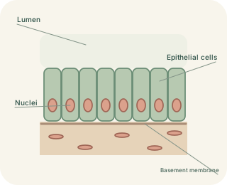
A continuous cellular boundary
covers surfaces and lines many tubes and cavities. Its cells fit closely together with relatively little extracellular material between them. An apical surface faces the outside or a , while the basal surface rests on a above supporting connective tissue. Cell junctions help control leaks and maintain attachment. Epithelia generally lack their own blood vessels, receiving nutrients across nearby tissue. This arrangement creates a controlled boundary rather than a loose collection of cells. Different boundaries need different combinations of thinness, strength and specialised transport.
Reading an epithelium
 Open diagram larger ↗
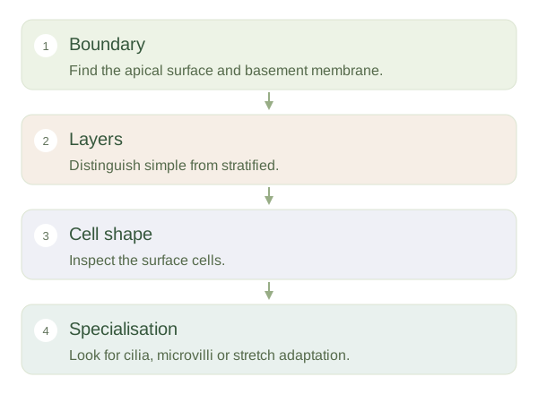
Count layers, then look at shape
Simple has one cell layer. Stratified epithelium has several layers and is named mainly for the shape of its surface cells. Squamous cells are flattened, cuboidal cells are roughly as tall as wide and columnar cells are taller. Simple squamous epithelium is suitable for rapid exchange, as in much of the gas-exchange surface. Stratified squamous epithelium resists abrasion in places such as the mouth and oesophagus. Simple columnar epithelium can support absorption and secretion in the intestine. Shape is a clue to function, not a complete description of it.
Special appearances need context
Pseudostratified looks multilayered because nuclei lie at different heights, although its cells contact the . Transitional epithelium, or urothelium, changes its surface appearance as parts of the urinary tract stretch. Cilia can move material along a surface, while microvilli increase the available membrane area. A tilted section can make one layer look thicker, so always examine the boundary and neighbouring regions. The practical approach is to identify the surface, count meaningful layers and then relate the pattern to the mechanical and transport demands of that organ.