Crystals in a concentrating system
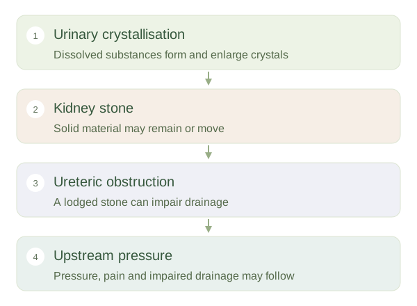
Kidney stones form when substances in urine crystallise and grow into solid material. Calcium oxalate is common, but stones can have other compositions, including uric acid, struvite or cystine. The presence of calcium in a stone does not mean dietary calcium is automatically its cause. Risk depends on the concentration of several substances, urine chemistry, inhibitors of crystal growth and individual conditions. Urine normally concentrates waste products, so stone formation reflects a balance between dissolved material remaining in solution and crystals developing.
How a stone can affect drainage
 Open diagram larger ↗
Why movement can produce pain
A stone may remain within a kidney without causing obvious symptoms. If it enters a and obstructs flow, pressure and ureteric muscle activity can cause severe pain. Pain may travel from the towards the groin as the affected location changes. Blood can appear in urine when the lining is irritated, but neither pain nor blood identifies a stone with certainty. The size, shape and location of a stone influence whether it passes and how much occurs; pain intensity alone does not measure stone size.
Obstruction links anatomy with risk
Urine must travel from collecting spaces through ureters into the bladder. An can raise upstream pressure and impair drainage. Infection combined with an obstructed urinary tract is particularly concerning because infected material cannot drain normally. Diagnosis may use urine testing and imaging to identify location and consequences, rather than symptoms alone. Different stone types have different risk factors, so prevention and treatment are individual clinical decisions. The educational principle is that a small solid object can have a large effect when it lodges in a narrow conducting tube. Stones demonstrate why fluid chemistry and regional anatomy must be understood together.